AR Liver Viewer

Description
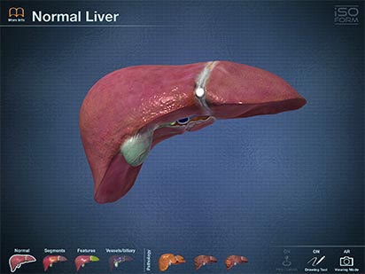
AR Liver is a real-time 3D medical education and patient communication tool, featuring incredibly detailed anatomical models. It is a member of a series of apps developed specifically for the iPad 2 and the New iPad using actual human CT imaging data, and the most accurate 3D modeling technology available.
AR Liver is appropriate for use by secondary students, undergraduate and graduate students, and medical professionals.
Full functionality of the app requires the downloading of a graphic known as a glyph that will be used during the Augmented Reality (AR) viewing experience.
Features
There are two ways to interact with AR Liver.
First, AR Liver utilizes true real-time 3D. Unlike other anatomical apps and programs, there are no pre-rendered frames or animations. Therefore, the user can place the incredibly detailed organ in any position and zoom in to any location to explore all of the anatomical structures. The user can learn by using the extensively labeled sections, study by creating custom labeled pins, or teach by drawing directly on the liver in 3D.
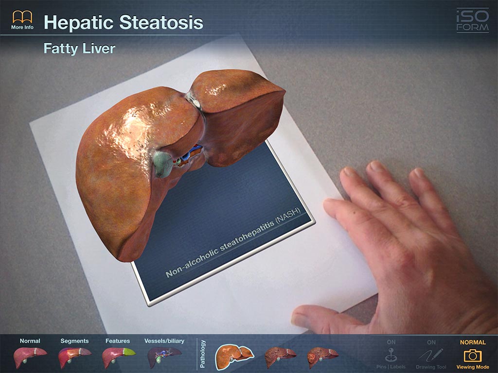
Second, AR Liver offers an Augmented Reality (AR) viewing mode. This mode bridges the gap between the purely computer based interactive experience and the real world. With the use of the iPad's camera and a printed graphic, known as a glyph, the user can view a virtual liver in any location as if it were sitting in front of them. By rotating the glyph or the iPad, the user can explore the external features of the organ.The user must download the printable graphic in order to view the AR function included in this app. Please visit
www.iso-form.com/AR.pdf to download the printable graphic.
Views:
By selecting the different views at the bottom of the interface the user can explore the liver using a series of optional views. Color-coded, didactic models help to show the specific locations and margins of the liver segments, while a semi-transparent view allows for the exploration of internal structures and features, including the major vessels and biliary tree. Finally, real-time 3D models of three common pathologies can be viewed to illustrate the anatomical changes that occur to the organ.
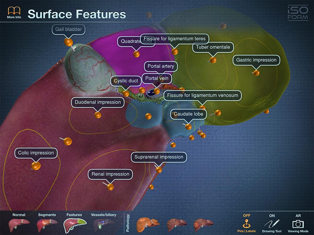
Labeling Pins:
By turning on the colored labeling pins, the user can study the names and locations of the anatomical structures, including: the segments, ligaments of the liver and various landmarks of other visceral organ impressions. The pins and labels remain on screen and in the exact anatomical location during all real-time 3D user interaction.
Pen tool:
Drawing with the multi-colored pen tool allows the user to draw on the surface of the 3D liver in order to highlight features, locations, and anatomical structures. Perfect for lectures, labs, or study groups.
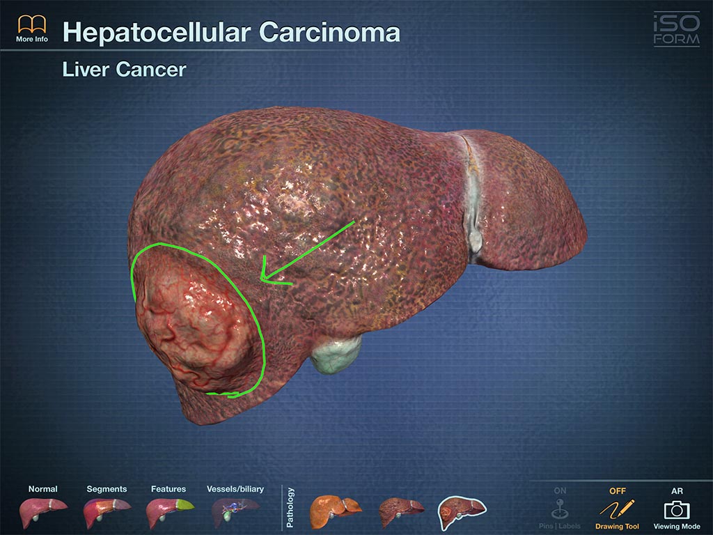
Pathologies:
AR Liver contains fully interactive real-time 3D models of three common hepatic pathologies: Hepatocellular Carcinoma (HCC), Cirrhosis, and Non-Alcoholic Steatohepatitis (NASH). Each of these detailed models includes a short description of the pathology shown.
Screenshots
Support
For any questions, comments, suggestions, or technical support requests, please email us at: contact@iso-form.com